
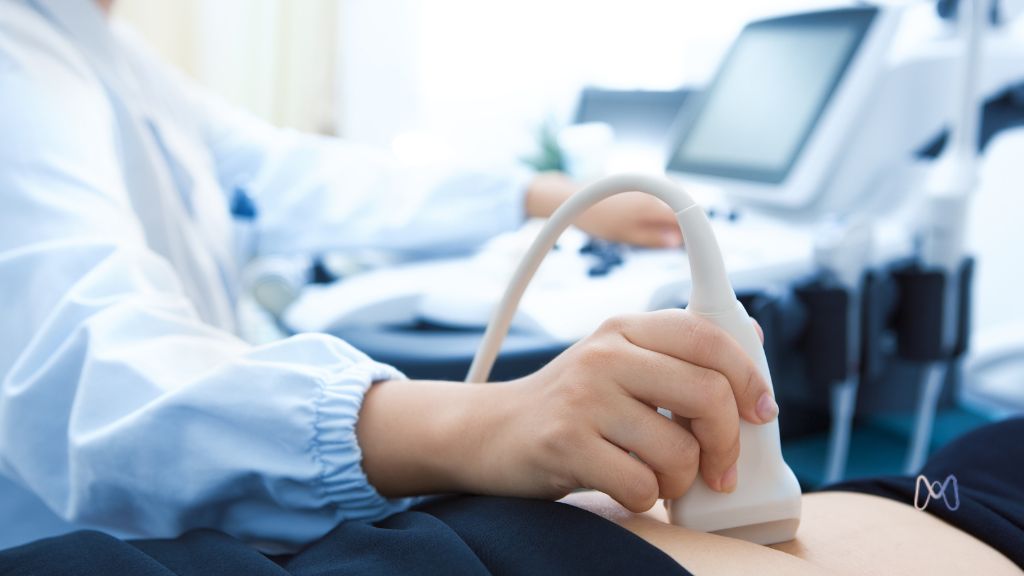
When a patient is asked to have an ultrasound with endometriosis mapping, one question often comes up: how is this exam different from the transvaginal ultrasound she may have had before?
The difference is not only in the equipment used. Ultrasound with mapping is directed at searching for signs of the disease in different pelvic regions. It follows a specific protocol, assesses organ mobility, looks for changes that may suggest adhesions and describes the probable location and extent of lesions.
Rather than simply answering whether endometriosis is present, the goal is to build an organized view of the pelvis and produce information that helps decide how to monitor or treat the patient.
What does it mean to map endometriosis?
Mapping means systematically searching for changes in regions where endometriosis can develop. In a conventional gynecological ultrasound, the assessment usually focuses on the uterus, endometrium, ovaries and more evident pelvic changes. With mapping, the examiner broadens this investigation.
- Uterus and ovaries
- Area behind the cervix
- Uterosacral ligaments
- Vaginal fornix
- Space between vagina and rectum
- Rectum and sigmoid colon
- Bladder and pelvic ureters
- Mobility between organs
- Possible signs of adhesions
One woman may have an ovarian endometrioma. Another may have a bowel nodule. Another may have deep changes behind the uterus. There are also patients with suggestive symptoms and no identifiable lesions on imaging. The exam must be interpreted within this diversity.
What is the difference between a regular ultrasound and mapping?
The transvaginal approach may be similar, but the purpose, protocol and depth of evaluation are different. A conventional ultrasound evaluates the uterus, endometrium, ovaries, fibroids, cysts and other gynecological conditions. Mapping also looks for signs of deep disease, involvement of other organs and reduced mobility of pelvic structures.
For this reason, it is not enough to add the phrase endometriosis assessment to any ultrasound. The result depends on an adequate protocol, time dedicated to the assessment, knowledge of disease presentations, examiner experience and an organized report.
Why may bowel preparation be requested?
Gas and residue can make it harder to visualize the bowel wall and regions located behind the uterus. Preparation aims to reduce this interference. Depending on the service, it may involve dietary instructions, laxatives or other measures.
The protocol is not the same in all institutions. The patient should follow the instructions from the place where the exam will be performed and should not adapt a preparation found online on her own.
Can the exam identify ovarian endometriosis?
Ultrasound can identify findings compatible with endometriomas, cysts usually filled with old blood content. The specialist evaluates size, location, internal appearance, relationship with ovarian tissue, mobility and association with signs of deep disease.
Identifying an endometrioma does not automatically determine surgery. Age, symptoms, desire for pregnancy, ovarian reserve, size and lesion characteristics must all be part of the decision.
Is it possible to identify bowel endometriosis?
Specialized ultrasound can identify nodules affecting the wall of the rectum or sigmoid colon. The report may describe location, size, extent, apparently involved layers, percentage of circumference involved and signs of reduced bowel caliber.
This information influences planning, but the exam does not decide by itself whether the patient needs surgery.
Can ultrasound assess the bladder and ureters?
Yes. Mapping can look for changes compatible with endometriosis in the bladder and observe signs related to the ureters, the channels that carry urine from the kidneys to the bladder. Deep endometriosis or fibrosis can narrow these channels and affect urine flow, not always with intense symptoms.
When urinary involvement is suspected, other exams may be necessary to complement assessment of the ureters and kidneys.
How does the exam identify possible adhesions?
Ultrasound does not show all adhesions directly, but it allows the examiner to observe how the organs move. Reduced mobility of the uterus, ovaries or bowel may suggest that structures are fixed.
This dynamic assessment is an important feature of the method. It does not record only a static image: it allows observation of the pelvis in motion and shows how organs relate to one another.
Why does examiner experience matter so much?
The professional moves the transducer, changes angles, applies controlled pressure, follows organ displacement and decides which regions require more attention. For this reason, two assessments performed with the same machine may produce different results.
- Recognition of lesions
- Assessment of harder-to-see locations
- Interpretation of organ mobility
- Differentiation between similar changes
- Quality of measurements
- Organization of a report that is useful for treatment
How did Brazil contribute to the evolution of this exam?
The development of ultrasound for mapping deep endometriosis includes an important contribution from Brazilian researchers. Dr. Maurício Simões Abrão reports that his search began with a question: why would it be necessary to operate on a patient only to understand where the disease was?
In collaboration with radiologist Dr. Manoel Orlando Gonçalves and other professionals, the team began correlating what appeared on ultrasound with what was found during surgeries. This connection helped develop and refine the protocol.
In 2007, a study led by Dr. Maurício compared clinical examination, transvaginal ultrasound and MRI in the evaluation of retrocervical and rectosigmoid deep endometriosis. The work showed the value of specialized ultrasound in preoperative planning.
Is ultrasound better than MRI?
There is no single answer, because both exams can identify and assess the extent of deep endometriosis when performed with adequate technique and interpreted by experienced professionals.
In our service, specialized transvaginal ultrasound with bowel preparation is the first-choice exam for investigating deep endometriosis. In addition to high diagnostic accuracy, it offers a dynamic pelvic assessment, allows observation of organ mobility, correlates painful points reported by the patient with the structures examined and is usually more accessible.
MRI remains an extremely important exam, especially when there is a need to complement investigation, assess specific locations or answer clinical questions that ultrasound alone does not clarify. It also offers a broad view of the pelvis and allows later review of images.
More important than choosing which exam is better is ensuring that the assessment is guided by the patient's clinical history and performed by teams experienced in endometriosis diagnosis.
Does a normal result rule out endometriosis?
No. Endometriomas and several deep forms can be identified, but superficial peritoneal lesions may not be visible. When the exam is normal, symptoms, relationship with the cycle, protocol quality, professional experience and other possible diagnoses must be considered.
The exam is part of the reasoning. It does not replace the reasoning.
Does lesion size show disease intensity?
The exam measures anatomical changes, but it does not measure suffering. A small lesion may be located in a sensitive region and produce important symptoms. A larger lesion may be found in a patient with few complaints. Inflammation, fibrosis, adhesions, nervous system sensitization and time living with pain also shape the experience.
Does mapping define whether surgery will be necessary?
No. The exam may show information that makes surgery more or less likely, but the indication also considers symptoms, impact on quality of life, organ involvement, response to clinical treatment, fertility, ovarian reserve, previous surgeries, risks and the patient's preferences.
How does the exam help when surgery is indicated?
Mapping can show ovarian, bowel, bladder, ureteral or vaginal involvement in advance. This helps estimate complexity, bring in professionals from other specialties, organize hospital resources and discuss risks and possible limits with the patient.
Does the exam usually cause pain?
Discomfort varies. Patients with pelvic pain, deep pain during intercourse, pelvic floor tension or lesions in certain regions may feel greater sensitivity. The patient should report pain so pressure and technique can be adapted.
When the transvaginal approach is not indicated, not accepted or not possible, other strategies, such as transabdominal ultrasound or MRI, may be discussed.
How should the patient prepare for the exam?
Preparation depends on the service protocol. It may involve a specific diet, medication to reduce gas, bowel preparation, fasting, bladder instructions and bringing previous exams.
- Previous ultrasounds and MRIs
- Surgical reports
- Pathology results
- List of treatments used
- Information about the menstrual cycle
- Description of the main symptoms
Conclusion
Ultrasound with endometriosis mapping is a specialized assessment that seeks to identify and locate changes in different pelvic regions. It can recognize endometriomas, deep forms, bowel or urinary involvement and indirect signs of adhesions.
Its value does not depend only on the equipment. It depends on the protocol, the examiner's experience and the ability to transform images into useful information. A positive result does not determine surgery, and a normal result does not exclude all forms of the disease.
More than searching for lesions, the exam helps transform a disease often discovered only during surgery into a condition that can be studied and planned in advance.
Frequently asked questions about ultrasound with endometriosis mapping
What is ultrasound with endometriosis mapping?
It is a specialized ultrasound that systematically evaluates different pelvic regions in search of endometriomas, deep endometriosis, organ involvement and indirect signs of adhesions.
What is the difference between a regular ultrasound and endometriosis mapping?
A conventional ultrasound mainly evaluates the uterus, endometrium and ovaries. Mapping uses a targeted protocol to investigate regions that may be affected by endometriosis and describe its probable extent.
Does ultrasound with mapping require bowel preparation?
In some services, yes. Preparation may reduce gas and residue that make it harder to visualize the bowel and deep pelvic structures. Instructions vary and should be provided by the clinic where the exam will be performed.
Can ultrasound identify bowel endometriosis?
It can identify nodules in the rectum or sigmoid colon and provide information about location, size and probable extent. Performance depends on the type of lesion, protocol and examiner experience.
Can ultrasound identify ovarian endometriosis?
Yes. It can identify findings compatible with endometriomas and assess size, location, appearance, ovarian mobility and association with deep disease.
Does a normal ultrasound rule out endometriosis?
No. Endometriomas and several deep forms may be identified, but superficial lesions may not appear. The result must be interpreted together with symptoms and clinical assessment.
Does ultrasound with mapping replace MRI?
Not in every case. The exams may be alternatives or complementary. The choice depends on the clinical suspicion, the possible lesion location and the expertise available.
Can mapping say whether surgery will be necessary?
Not by itself. Surgical indication considers symptoms, involved organs, response to treatments, fertility, risks and the patient's goals.
Why is examiner experience important?
Because ultrasound is dynamic and depends on how the professional searches for, recognizes, measures and interprets the findings.